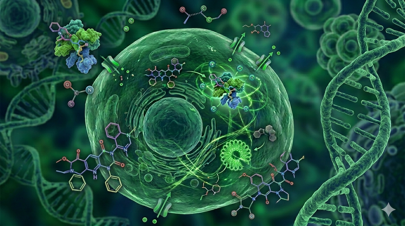
Biochemistry can tell you what a molecule does to a purified protein in a test tube. What it cannot tell you is whether that molecule will do anything useful inside a living cell. Cells are dynamic systems with membranes, transporters, metabolic enzymes, and competing targets that can redirect a compound’s activity in ways no cell-free experiment can predict. That gap is what cell-based assays are designed to bridge.
Today, cell-based assays for drug discovery are used at nearly every stage of the research pipeline – from the first round of compound screening to preclinical safety testing. Understanding what they are and how they work is useful not just for bench scientists but for anyone involved in structuring a drug discovery program.
What Is a Cell-Based Assay?
A cell-based assay is an experimental measurement that uses living cells as the biological system. Rather than isolating a single enzyme or receptor and measuring its behavior in solution, a cellular assay exposes intact cells to the compound of interest and measures their response – whether they survive, proliferate, activate a signaling pathway, express a reporter gene, change shape, etc.
The cells used can vary widely: established cancer cell lines, primary cells from human or animal tissue, stem cell-derived models, or engineered lines expressing a specific target. Each offers different trade-offs among biological relevance, experimental consistency, and practical accessibility.
What distinguishes cellular formats from biochemical ones is context. A compound has to cross the cell membrane to reach an intracellular target. Once inside, it encounters metabolizing enzymes that may inactivate it, efflux pumps that may expel it, and off-target proteins that may bind it before it reaches the intended site. All of these factors are invisible in a biochemical assay and fully present in a cell-based one, which is what makes cellular measurements harder to run and more informative when they work.
Why Utilize Cell-Based Assays?
Drugs work in organisms, not in solution. At some point in the development process, a compound has to prove that it can do something meaningful in cells before anyone will invest in animal studies or clinical trials. Cell-based experiments are the earliest systematic point at which that proof can be generated.
Beyond relevance, live-cell assays offer something biochemical formats cannot: the ability to observe a compound’s effect on a functional biological process rather than just a molecular interaction. A kinase inhibitor may bind its target with high affinity in a cell-free system, but if it fails to penetrate the membrane or if the pathway has redundant inputs that compensate for the block, its biochemical activity is largely irrelevant to its potential as a drug.
Cell-based formats also provide early toxicity data. Cytotoxicity and mitochondrial function assays can flag compounds that damage cells at concentrations close to their active dose – a signal that almost always predicts problems further down the pipeline. Catching it before multiple chemistry cycles have been invested in a compound saves significant time and resources.
Some biological targets cannot be addressed at all in a biochemical format. Membrane-bound receptors, ion channels, and multi-subunit complexes often lose their functional properties when extracted from the cellular environment. For these targets, a cell-based experiment is not just preferable – it is the only practical option.
Why Cell-Based Assays Matter in Drug Discovery
Drug discovery programs fail most often not because the chemistry is wrong, but because the biology turns out to be more complicated than early experiments suggested. A compound that appears to be a clean inhibitor in a biochemical screen may exert its effects through a different mechanism of action in cells, or may activate compensatory pathways that neutralize its intended effect. Cell-based assays are the earliest opportunity to discover these discrepancies before resources are committed to the wrong candidate.
In high-throughput screening, cell-based formats are widely used for primary screens. This reflects a broader shift toward phenotypic approaches that ask what compounds do to cells rather than what they do to a single isolated protein. Phenotypic screens have historically contributed many clinically successful drugs, especially where the target biology was not fully defined.
Cell-based data also feeds directly into lead optimization. When medicinal chemists modify a compound’s structure to improve potency or selectivity, they need to know whether those modifications translate into improved cellular activity. A compound that gains potency in a biochemical assay but loses cell permeability due to a structural change has not improved in any clinically meaningful way. Cellular measurements keep optimization grounded in biological reality.
Common Types of Cell-Based Assays
The range of formats in use today reflects the diversity of questions that drug discovery programs must address.
Cell viability and cytotoxicity assays are among the most widely used. They measure whether cells remain alive and metabolically active after compound exposure, typically using colorimetric reagents such as MTT or resazurin, or luminescent ATP detection. These assays provide a direct measure of compound toxicity and are routinely run in parallel with activity assays to establish a therapeutic window.
Reporter gene assays link a signaling pathway to the expression of a detectable protein, most commonly luciferase or a fluorescent marker. When a compound activates or inhibits the pathway, reporter expression changes, producing a quantifiable signal. These formats are well-suited for screening modulators of transcription factors, nuclear receptors, and other transcriptional regulators.
Calcium flux assays measure rapid changes in intracellular calcium concentration, which serve as second messengers for GPCRs, voltage-gated ion channels, and receptor tyrosine kinases. This format is broadly applicable for target classes that are difficult to address in purely biochemical systems.
High-content imaging assays capture detailed phenotypic information from cells using automated fluorescence microscopy. Instead of reducing cellular behavior to a single number, these formats extract dozens of measurements per image – nuclear morphology, cytoskeletal organization, protein localization – to build a multi-dimensional profile of compound effects. They are increasingly used for mechanism of action studies and for detecting off-target effects that simpler assays would miss.
Three-dimensional culture models, including spheroids and organoids, allow cells to grow in architectures that more closely resemble actual tissues. They are especially valuable in oncology and fibrosis research, where tumor microenvironment and tissue architecture strongly influence drug response.
How Cell-Based Assay Development Works
Running a cell-based assay may seem straightforward, but designing one that is robust and reproducible is considerably more complex. Assay development defines and optimizes all parameters that determine how the assay behaves – and it typically precedes any compound screening by weeks or months.
The first decision is cell line selection. The chosen cells need to express the target at relevant levels and in a form that reflects the biology the assay is meant to model. For some targets, recombinant overexpression in HEK293 or CHO cells is appropriate. For others, where expression level or post-translational modification matters, primary or disease-relevant cells are needed.
Once a cell system is selected, assay optimization addresses practical parameters such as seeding density, incubation times, reagent concentrations, solvent tolerance, and plate format. Each variable can shift the assay window – the separation between the maximum and minimum signals. The Z’-factor, calculated from control well variability, benchmarks this separation quantitatively; a Z’ above 0.5 is generally required before a format is considered ready for screening.
Assay validation confirms that the assay measures what it claims to measure, that results are consistent across plates and operators, and that known actives and inactives are correctly identified. Validation continues through the life of a campaign, with control data tracked on every plate to detect performance drift early.
Benefits and Challenges of Cell-Based Assays
The central benefit of cell-based assays for drug discovery is their biological relevance. Beyond that, these formats can measure multiple endpoints simultaneously – cytotoxicity alongside target engagement, or pathway activation alongside phenotypic change. They scale to 384- and 1536-well plates for high-throughput use and, in kinetic mode, capture time-resolved data on the dynamics of compound effects.
The challenges are real. Cell-based assays are more variable than biochemical ones. Cells change with passage number, culture conditions, and reagent lot variation. This variability can be controlled through careful cell banking and quality monitoring, but it requires sustained effort and cannot be eliminated entirely.
Compound interference is a persistent concern. Some compounds fluoresce at assay wavelengths or aggregate under standard conditions, leading to non-specific apparent inhibition. Counter-screens and orthogonal formats are standard tools for filtering these artifacts.
Interpreting results from complex cell-based formats also requires genuine biological expertise. Understanding why a compound alters nuclear morphology or why reporter expression increases in one cell line but not another requires familiarity with signaling pathways, cell biology, and assay design principles. This expertise is embedded in experienced research teams and in well-structured, integrated drug discovery services that combine assay execution with scientific interpretation.
Despite the challenges, the field is clearly moving toward earlier cell-based data throughout the process. The high failure rate of compounds in clinical trials – largely attributed to insufficient biological characterization in early discovery – has made hit identification in cellular systems the expected standard rather than an optional step. Programs that invest in rigorous assay development, proper assay validation, and careful result interpretation consistently produce better-characterized compounds with a higher probability of surviving the later stages of development.
Thus, cell-based assays are where promising chemistry meets biological reality. By revealing whether a compound can truly modulate cellular pathways without unacceptable toxicity, they help transform early hits into drug candidates that are worth advancing.